Patient Communication
Why dental radiographs are often hard to explain to patients
Looking at the same image does not mean understanding it in the same way.
Redazione Vesalya · September 17, 2026 · 6 min read
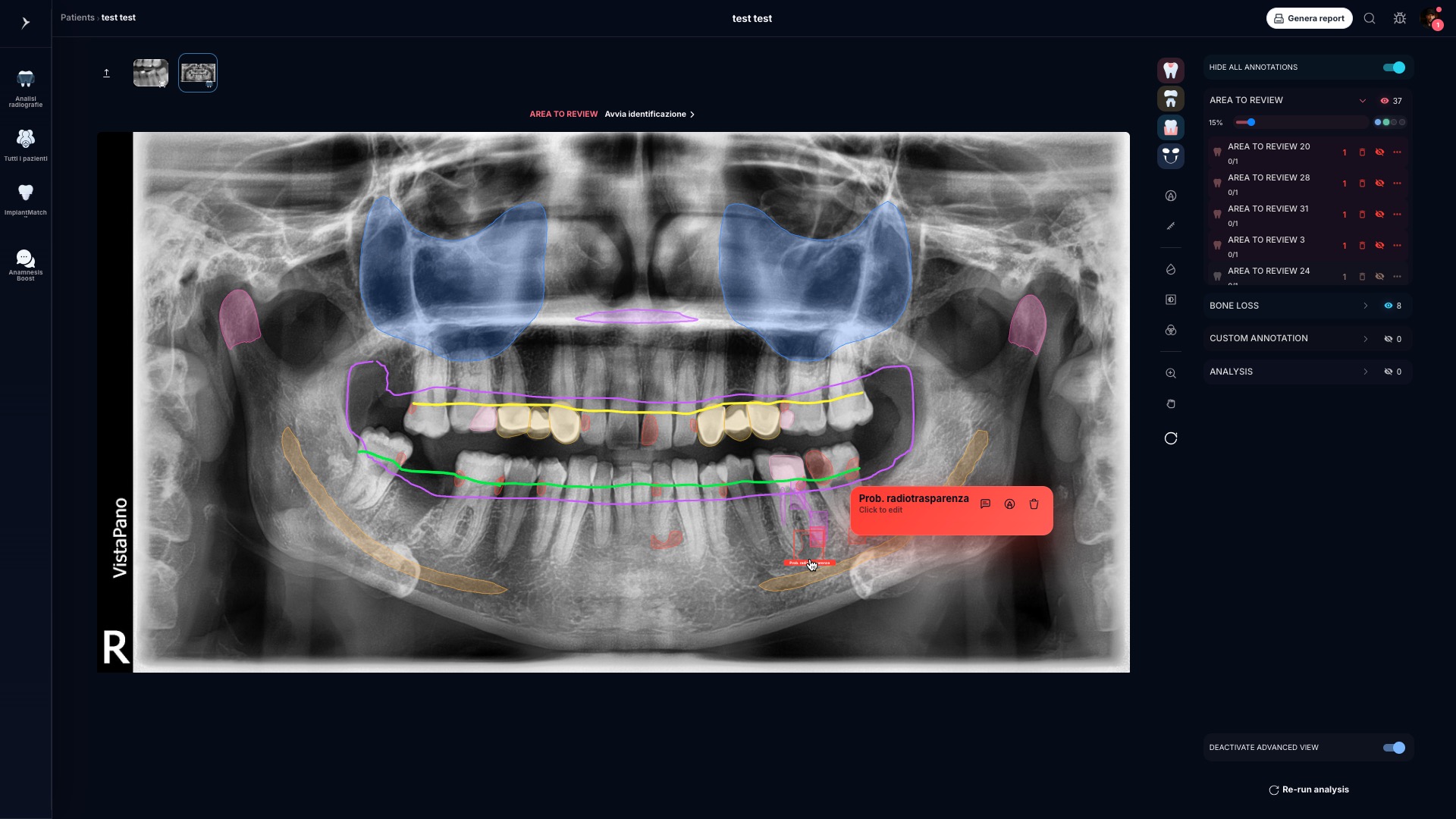
The dentist opens the radiograph on the monitor, points to a specific spot and says something that sounds simple: “Look here.” To the dentist, it is obvious where to look and why that area deserves attention. The patient leans towards the screen, studies the image and often nods without being sure they have really understood.
It is not a lack of attention, and it is not necessarily a communication problem on the professional’s side. Dentist and patient are looking at the same image, but they are not seeing it in the same way.
For the dentist, a radiograph is a set of information to be read in its clinical context. For the patient, it may be nothing more than a complex arrangement of whites, blacks and shades of grey, with no immediate points of reference.
The dentist does not just look: they interpret
When a professional looks at a radiograph, they do not start from scratch. Training and experience allow them to recognise anatomical structures, restorations, crowns, implants, endodontic treatments and variations in the image that may call for a closer look.
Above all, what they see is never considered in isolation. The radiograph is related to the medical history, the reported symptoms, the clinical examination, previous treatments and the other information available. It is the professional who gives the image its meaning within the case.
The patient does not have the same points of reference. They cannot necessarily tell an anatomical structure from an existing treatment, or recognise which difference is relevant and which is part of the normal variability of the image. Before they can even understand the explanation, they need to know where to look.
“Can you see this darker area?”
It is one of the most common phrases during an explanation. The dentist immediately identifies the area and compares it with the surrounding structures. The patient, on the other hand, may not understand which of the many dark areas in the image is the one being pointed at.
The same happens with phrases such as “look at this margin” or “compare this area with the one on the other side.” They are precise instructions for someone used to reading radiographic images; for someone who only sees them during a visit, they require considerable effort.
The difficulty therefore arises before the words. If the patient cannot confidently identify the area being discussed, even a clinically correct explanation risks remaining abstract.
Explaining more does not always mean explaining better
When an explanation does not seem to land, the most natural reaction is to add detail: more information, more terms, more steps. But a radiograph already contains a great deal of visual stimuli.
During the visit, the patient has to orient themselves in the image, distinguish structures they do not know, follow the professional’s language and connect what they are hearing to the possible consequences for their own care pathway, all at the same time. Increasing the amount of information can also increase the cognitive load.
The useful question, then, changes. Not only: “How can I explain this radiograph?” But: “How can I help the patient see what I am explaining?”
First guide the eye, then build the explanation
A clearer conversation can start with a simple sequence:
- define the part of the image you want to talk about;
- visually guide attention towards the right area;
- explain, in accessible words, what the professional is observing;
- connect the observation to the clinical picture and the care pathway;
- check understanding and leave room for questions.
The goal is not to teach the patient to read a radiograph on their own. Interpretation, assessment and decision remain the professional’s responsibility. The goal is to allow the patient to better follow the reasoning the dentist is sharing.
Simplify the language without trivialising it
The gap is not only visual. Clinical terminology allows professionals to communicate precisely, but it can feel unfamiliar to those who do not use it every day.
Simplifying does not mean making the information less accurate or turning a complex assessment into an absolute answer. It means choosing understandable words, introducing one concept at a time and linking it to a recognisable visual reference.
When the dentist can say “this is the area we are talking about” and the patient can clearly identify it, image and language start working together. The patient no longer has to imagine what the professional is describing: they can observe it together.
From trust to shared understanding
Trust remains central to the relationship between dentist and patient. A visual explanation does not replace it: it can strengthen it.
Saying “trust me” asks the patient to accept a conclusion. Saying “look here” — when what is shown is genuinely clear — makes it possible to build the conversation around a common reference. The professional continues to interpret; the patient can better understand the reason for the explanation and ask more relevant questions.
This does not mean that all uncertainty disappears, or that an image alone can explain the whole case. It means creating the conditions for a more transparent conversation, in which the patient can take part with greater awareness.
Visual communication is not self-diagnosis
Making a radiograph easier to read does not mean giving it an automatic meaning, nor inviting the patient to draw clinical conclusions on their own.
Highlighting an area, placing two images side by side or organising the information can make the explanation easier. The meaning of what is shown, however, depends on the clinical context and on the professional’s assessment.
The radiograph is only one step of the pathway
The conversation does not necessarily end in front of the monitor. New questions may arise after the visit; some details may be forgotten; the treatment plan may require a later discussion with the patient or with other members of the team.
This is why it helps to see the radiograph as part of a broader flow:
The clarity built during the visit becomes more valuable when it can continue afterwards: in the information shared with the patient, in the case documentation and in the continuity of communication within the practice.
What role can technology play?
Digital tools can help organise and visualise the information contained in radiographic images more effectively. Artificial intelligence can also offer support, provided its role remains defined and supervised.
It should not turn a radiograph into an automatic answer, replace the dentist’s interpretation or create shortcuts in the clinical process. It can, however, help highlight information for the professional to review and make its communication more orderly.
The principle remains simple: artificial intelligence supports the analysis; the professional reviews, interprets and decides.
Where Vesalya fits in
Vesalya was created in the space that connects radiographic review, patient explanation and case documentation.
The platform is designed to support dentists in making radiographic information clearer, more visual and more structured throughout the practice workflow, always keeping the professional at the centre of the assessment.
Not to replace clinical judgement, but to ease the step between what the dentist observes, what they choose to explain and what they need to document.
Because a radiograph can contain a great deal of information. Its value in the relationship with the patient also depends on the ability to turn that information into an understandable conversation.
In summary
- Dentist and patient look at the same radiograph with different visual references.
- An effective explanation starts by guiding the eye, not by adding technical terms.
- Visual clarity and accessible language can reinforce each other.
- Interpretation and clinical decisions always remain the professional’s responsibility.
- Technology and AI can support visualisation, organisation and documentation, under clinical supervision.
To learn about Vesalya’s approach to radiographic review, patient communication and case documentation, explore the Vesalya workflow.